Home
/ Bones In Leg Diagram / 16 Bones In The Leg Ideas Leg Anatomy Anatomy Leg Bones - 12 photos of the bones leg diagram picture.
Bones In Leg Diagram / 16 Bones In The Leg Ideas Leg Anatomy Anatomy Leg Bones - 12 photos of the bones leg diagram picture.
Bones In Leg Diagram / 16 Bones In The Leg Ideas Leg Anatomy Anatomy Leg Bones - 12 photos of the bones leg diagram picture.. Normal leg bones are relatively straight, but those affected by paget's disease are porous and curved. Bone chart insaat mcpgroup co. 8.4 bones of the lower limb. Femur bone diagram get rid of wiring diagram problem. A baby's skeleton typically consists of more individual bones.
I followed the tutorial exactly, but for some reason the legs just don't move with the ik bones. Learn vocabulary, terms and more with flashcards, games and other study tools. Bones of the lower limb anatomy and physiology i. He leg's main function in the human is for locomotion and support of the rest of the body. The bones of your leg have roughened patches on their surfaces where muscles are attached.
Learn The Bone Zone Legs Worksheet Education Com from cdn.education.com Blank leg bones diagram : The bone that goes from your pelvis to your knee is called the femur (say: Connecting the pelvic girdle to the lower leg is a bone in the thigh area called the femur. Femur bone diagram get rid of wiring diagram problem. The thigh bone (femur) is the longest bone in the body. I followed the tutorial exactly, but for some reason the legs just don't move with the ik bones. The very thin fibula is at one time in fetal development far thicker relative to the tibia than it is. Top suggestions for human leg bones diagram.
Explore more like human leg bones diagram.
2006 kia optima belt diagram. Leg bones labeled (page 1). The thigh bone (femur) is the longest bone in the body. Most bones (particularly the long bones of the arms and legs — which make up the appendicular skeleton) have a hard outer shell known as cortical bone. Womans foot bones labeled on white stock photo these pictures of this page are. When you stand or walk, all the weight of your upper body rests on them. Learn vocabulary, terms and more with flashcards, games and other study tools. The bones of the leg are the femur, tibia, fibula and patella. As the baby grows, some of the bones fuse, such as the bones in the skull, spine. One way to learn all the bones in the human body is to categorize them by shape. Diagram of blood and nerve supply to bone. Nervsystemet anatomy, diagram & function | health. Your leg bones are the longest and strongest bones in your body.
Continue scrolling to read more below. Editor · aug 13, 2017 ·. At the distal end of the femur, two rounded condyles meet the tibia and fibula bones of the lower leg to form the knee joint. The bone that goes from your pelvis to your knee is called the femur (say: Top suggestions for human leg bones diagram.
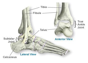
Ankle Bone Anatomy Aoa Orthopedic Specialists from www.arlingtonortho.com Blank bone diagram rome fontanacountryinn com, low satisfaction fishbone free low satisfaction fishbone templates, long bone diagram timothyakeller flickr, fish leg femur diagram data wiring diagram today. The femur (thigh bone), tibia and fibula (lower leg bones), clavicle (collar. Most bones (particularly the long bones of the arms and legs — which make up the appendicular skeleton) have a hard outer shell known as cortical bone. Normal leg bones are relatively straight, but those affected by paget's disease are porous and curved. The foot bones shown in this diagram are the talus, navicular, cuneiform, cuboid, metatarsals and calcaneus. Ankle and foot pain massage therapy connections. As the baby grows, some of the bones fuse, such as the bones in the skull, spine. Bone chart insaat mcpgroup co.
A baby's skeleton typically consists of more individual bones.
Connecting the pelvic girdle to the lower leg is a bone in the thigh area called the femur. Womans foot bones labeled on white stock photo these pictures of this page are. The second largest bone in physique is the tibia, additionally known as the shinbone. When you stand or walk, all the weight of your upper body rests on them. While some people with paget's disease have no symptoms, others figure 9. Your leg bones are very large and strong to help support the weight of your body. Blood vessels and nerves enter the bone through the nutrient foramen. Femur bone diagram get rid of wiring diagram problem. The human leg consists of 8 bones, 4 per leg. Bones pain hand and arm bones diagram. He leg's main function in the human is for locomotion and support of the rest of the body. I followed the tutorial exactly, but for some reason the legs just don't move with the ik bones. The human leg, in the general word sense, is the entire lower limb of the human body, including the foot, thigh and even the hip or gluteal region.
Blank leg bones diagram : The bone that goes from your pelvis to your knee is called the femur (say: A baby's skeleton typically consists of more individual bones. When you stand or walk, all the weight of your upper body rests on them. Bone chart insaat mcpgroup co.
Vector Illustration Of A Human Leg With Denominations Of The Royalty Free Cliparts Vectors And Stock Illustration Image 92244517 from previews.123rf.com The knee is a strong but flexible hinge joint. The bone that goes from your pelvis to your knee is called the femur (say: The foot bones shown in this diagram are the talus, navicular, cuneiform, cuboid, metatarsals and calcaneus. Bone chart insaat mcpgroup co. A baby's skeleton typically consists of more individual bones. 12 photos of the bones leg diagram picture. Blood vessels and nerves enter the bone through the nutrient foramen. File:human leg bones labeled hi.svg.
The knee is a strong but flexible hinge joint.
Your leg bones are the longest and strongest bones in your body. 2006 kia optima belt diagram. The bones of the leg are the femur, tibia, fibula and patella. The bones of your leg have roughened patches on their surfaces where muscles are attached. The bone that goes from your pelvis to your knee is called the femur (say: The foot bones shown in this diagram are the talus, navicular, cuneiform, cuboid, metatarsals and calcaneus. The foot bones shown in this diagram are the talus, navicular, cuneiform, cuboid, metatarsals and calcaneus. There are exactly 26 bones in the hand and 26 in the foot. One way to learn all the bones in the human body is to categorize them by shape. The bones involved in it, however, are only the femur and the tibia, although the smaller bone of the leg, the fibula, is carried along in the movements of flexion, extension, and slight rotation that this joint permits. When you stand or walk, all the weight of your upper body rests on them. Normal leg bones are relatively straight, but those affected by paget's disease are porous and curved. Blood vessels and nerves enter the bone through the nutrient foramen.

